 Eye Laser Clinic
Our clinic is located in the European part of Istanbul near the Bosphorus. Because of its location, it is easy to reach the Bosphorus, old and new districts, world famous museums and other historical monuments.
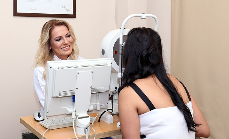
All examination equipment has cutting-edge technology and are maintained regularly. This equipment include the following:
Pentacam
50 different images of the eye illuminated with an interim light are obtained in a few seconds by way of rotation using a camera, which is kept inclined according to the Scheimpflug principle. Thus, 25.000 different values belonging to the anterior part of the eye are obtained. The examined part is between the cornea, iris and lens. All measurement values and images are recorded to make comparison with subsequent examination results. Printouts of these data can be obtained when necessary.
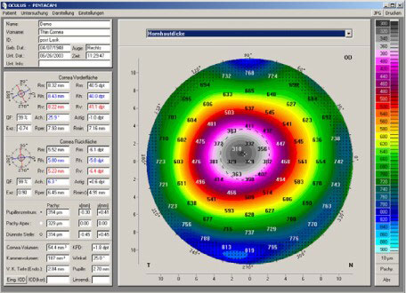
The opportunities provided by Pentacam:
- Image of large measurement results by way of 3-dimensional analysisMeasurement results include the following:
1) Chamber angle 2) Chamber volume 3) Chamber height 4) Central diameters
5) Astigmatism 6) Lens thickness - Pachymeter of the whole cornea
- The thickness of the cornea is determined over the whole area.
- Topography of the anterior and posterior areas of the cornea
- The state of the cornea is determined with height and curvature data.
- Interactive 3-dimensional picture of the eye part taken (to inform patients easily).
- Tomography shows the areas belonging to the anterior part of the eye as a 3-dimensional model in rotation.
- Cataract analysis with Densitogram and Scheimpflug shooting
- The cloudiness of the lens can be determined objectively by way of Densitogram and evaluated by calculating the precentages. The size of the cataract is determined manually by Scheimpflug shooting.
- Short measurement time (2 seconds) and automatic measurement assessment
Colvard Pupillometer
Colvard-Pupillometer provides the opportunity to measure the width of the pupil in the setting of daylight or twilight. This examination is very important before refractive surgery. With this result, the size of the area where treatment will be applied (Laser-ablation area where optic is used) is specified so that visual ability is not limited or limited to a small extent in the twilight or night. The objective is to prevent undesirable conditions including halo.
Lensmeter
With the help of lensmeter the glasses of the eyeglasses used by the patient are measured automatically.
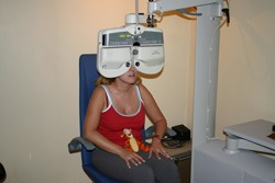
Auto-Refkeratometer
Auto-Refkeratometer has the property of automatic focusing and localization. With this device, mean ocular values can be determined. In addition, the corneal curvature is also measured.
Auto-Foropter
Oto-Foropter provides the opportunity of full measurement of the ocular values with automatic change of the lenses. The data of the Auto-refkeratometer and Lensmeter are transferred to the Foropter.
Automatic Tonometer
Automatic Tomometer has the property of automatic focusing and localization. It is tonometer that is used without contact. It does not contact with the cornea and therefore the risk of infection is completely eliminated. Intra-ocular pressure is measured with the help of tonometer.
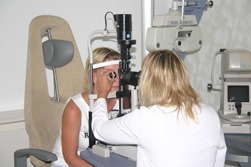 Biomicroscope
Different segments of the eye are examined with biomicroscope. In addition, the retina is checked with a special lens.