The Human Eye
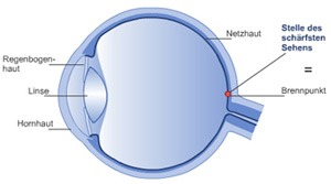
The eye can be compared to a camera. A camera consists of a lens system that can be adjusted by distance (Objective), a protector to adjust light, a film, a cover and a body. The eye also has a lens system. It is composed of a fixed anterior lens (cornea) and an adjustable lens (eye lens). The iris is the protector, and the retina is the film. The body of the eye is the skin part and is nourished by the vascular layer of the eye.
 Eyelids
The eyelids have various functions. When their structures are examined, their functions can be understood better. The eyelid is composed of two layers. The upper layer (including the skin of the eyelid) has two muscles. The first muscle (Musculus orbicularis oculi) is in the form of a circle around the eyelids, which is used to close the eyes. The second muscle (Musculus levator palpebrae) is used to open the eyes.
The second layer contains the eyelid cartilage (Tarsus). This tissue gives the eyelid its form and tightness. Below this, there is the conjunctival layer of the eyelid and covers the whole eye. The border between the two layers can be seen as a thin grey line at the edge of the eyelid when examined carefully.
The Meibomian glands extend through the cartilage of the eyelid up to the edge of the eyelid. The fluids they secrete constitute the fatty part of the tear. Chronic inflammation and fluid obstruction in these glands is called chalazion. There are also small glands in the eyelash part (glands of Moll and Zeis). Acute inflammation of these glands is called stye (hordeolum). Stye can also occur in the meibomian glands as an acute inflammation.
A small hole is observed at the edge of the upper and lower eyelids towards the internal part; these are teardrop duct holes. With each blinking of the eyelid tear is pumped from these holes towards the lacrimal canaliculi in the nose. With every blink, tear is reextended on the eye and thus the eye remains wet. Normally, this occurs unconsciously approximately 5-10 times a minute.
Tear
The lacrimal gland, which produces the majority of our tear, is localized in the external upper part of the eye. When the upper eyelid is elevated, the lower edge of the lacrimal gland is observed through the conjunctival layer. The outlet canaliculi of the lacrimal gland are also found here, but they are not visible. We produce approximately 3,5 milliliter tear fluid a day. Our tear is composed of 98% water and the remainder is composed of protein, salt and metabolism substances.
The tear stratum consists of three layers. There is a thin mucosa layer just on the eye (mucin), this is followed by an aqueous layer and this layer is covered with a thin adipose layer (lipid layer). This lipid layer prevents early evaporation of tear. The tear flows from the tear duct holes localized inside the upper and especially lower eyelid down towards the lacrimal canliculi. These small lacrimal canaliculi reach the lacrimal sac which is localized below the nasal bone.
Eyeball
The eyeball (Bulbus oculi) is found in the eyehole (Orbita), which is composed of many bones and has the shape of a funnel opening anteriorly. There is an adipose tissue covered with the conjunctival layer between the eyeball and the bone.
Six external muscles are found in the eyeball. Internal and external smooth muscles are found in both sides (Musculus rectus internus and Musculus rectus externus). Upper and lower smooth muscles are found above and below (Musculus rectus superior and Musculus rectus inferior). The upper oblique muscle (Musculus obliquus superior) extends towards the upper internal posterior part of the eye over a bony process in the form of a hook. The lower oblique muscle is found below the eyeball (Musculus obliquus inferior). The origins of the muscles are found in the depths of the eyeball. These six muscles are supplied by three nerves. The external ocular muscle has a special nerve (Nervus abducens) and the upper oblique muscle also has a special nerve (Nervus trochlearis); the other 4 muslces have a common nerve (Nervus oculomotorius).
The extraocular muscles are bound to the walls of the external conjunctival layer tissue of the eyeball (Sclera). The vessels and nerves nourish the eyeball by the sclera. The optic nerve (Nervus opticus) exits behind the eyeball through the sclera. It is composed of approximately 1,2 million nevre fibers and its total diameter is approximately 1,5 mm. It is surrounded by the meninx.
The eyeball (bulbus) is composed of three layers. The external layer – fibrous layer (tunica fibrosa) – is composed of the sclera and cornea. The inner layer- vascular layer (chorio idea)- is composed of the corpus ciliare and the iris – tunica vasculosa. The inner layer is composed of the retina.
Sclera
This is a fibrous layer and is the tightest tissue in the human body. It gives the eye its shape, and it protects the eye.
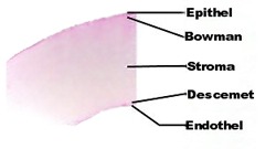
Cornea
The cornea is found on top of the sclera in the shape of a watch-glass and is bound to the sclera. It is transparent and sensitive to contact (protective measure). It constitutes a powerful area together with the water inside the eye chamber. This area constitutes approximately 75% of the refraction value of the eye. The lens and cornea work together to produce a clear image on the retina.
Fibrous layer (Choroidea)
This is the region of the eye with the most blood content. In addition, there is a lot of pigment (black spots) in the fibrous layer. It nourishes the eye excluding the retina and darkens the internal part of the eye.
Ciliar body (Corpus Ciliare)
It has the shape of a ring, containing glands in the creases on its surface, and is an elastic structure. The glands produce and release the chamber fluid. The accommodation muscle in the form of a ring is found inside the ciliar body. The function of the ciliar body is to produce the chamber fluid, to flatten the eye lens and to provide accommodation.
Iris
The iris has the shape of a round disc. The pupil is found in the middle of the iris. The pupil moves with two muscles. As age advances, the pupil loses its ability to move. By adjusting the amount of light entering the eye, the iris protects it.
The eye lens (Lens cristallina)
The eye lens contains fluid, which intensifies towards the center. It is inhomogeneous optically. It does not contain vessels or nerves. It is responsible for focusing light rays on the retina.
The Vitreous Body (Corpus Vitreum)
The vitreous body is a gelatinous substance ( 98% water). It is covered by a thin membrane. It has no nerves or vessels. Cloudiness and bands occuring in the vitreous body may form an image of shadow on the retina.
Retina
The retina is a transparent membrane. Generally, it is composed of receptors- cone cells and rod cells. Both are sensory cells and have cross-sectional areas with 6 corners. The cone and rod cells transform the light into a nerve stimulant.
Cone cells
We see colors, figures and movements with the cone cells. These cells are more sensitive to light. Therefore, we can only see during the daytime with them. (In the daylight we can see clearly and we can see the colors.)
Rod cells
With these cells we see coarse forms and movements in the twilight. We see colorless and unclear figures with them in settings with low light.
Blind spot (Papille)
Blind spot is where the optic nerve fibers exit the eye. The rod and cone cells are not present here; therefore, this place is named the blind spot.